Osteodystrophy & Growth Plate Abnormalities in Children: Causes, Diagnosis & Treatment
Quick Takeaways
- Osteodystrophy covers several metabolic bone disorders that hinder normal growth in kids.
- Growth‑plate (physis) abnormalities often signal an underlying systemic problem.
- Key labs: calcium, phosphate, alkaline phosphatase, PTH, vitamin D.
- Imaging-X‑ray and MRI-pinpoint structural changes early.
- Management blends nutrition, medication, and, when needed, orthopedic surgery.
Osteodystrophy is a group of metabolic bone disorders that disrupt normal mineralization and growth in children. It can stem from renal failure, nutritional deficits, or genetic mutations, and each cause leaves a distinct pattern on labs and X‑rays.
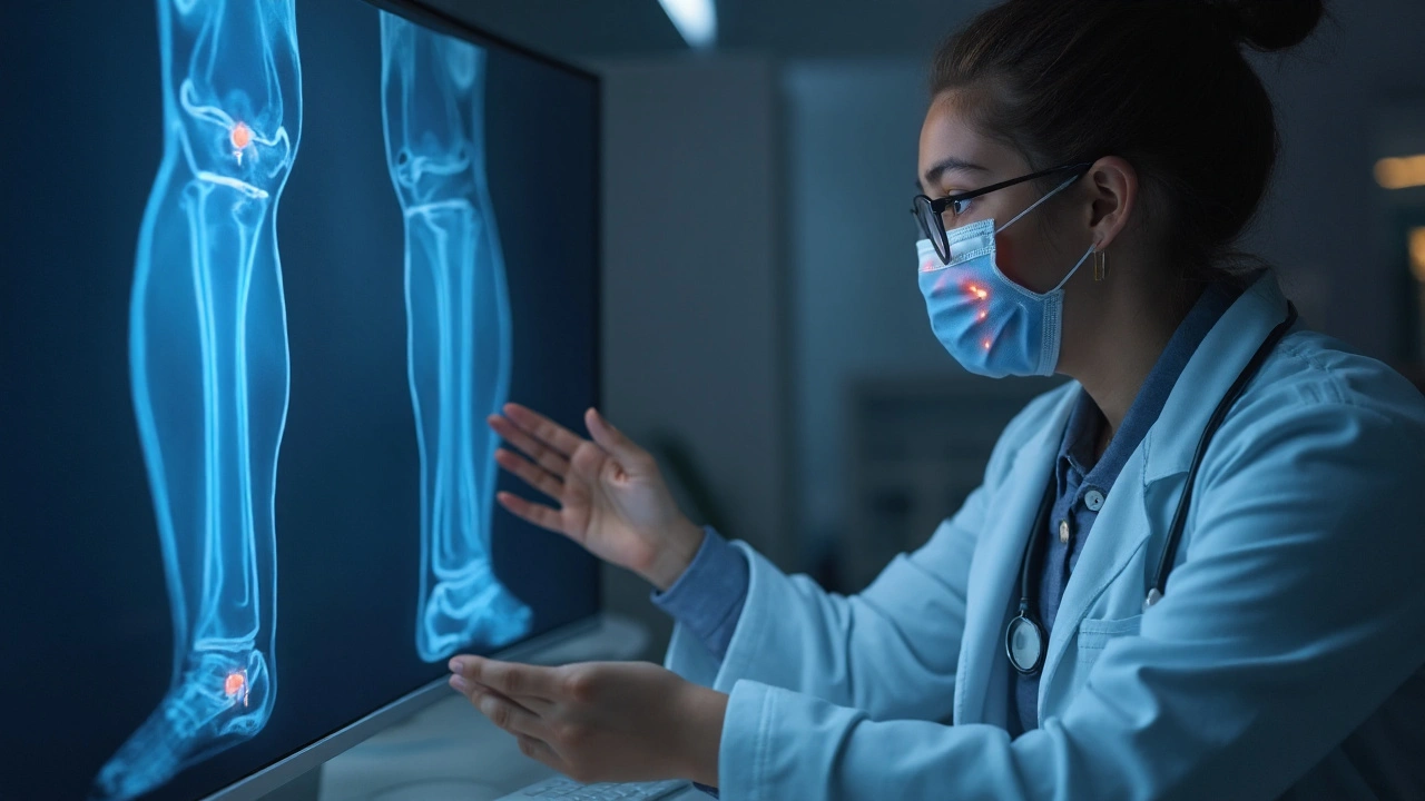
Growth Plate (also called the physis) is the cartilage region at the ends of long bones where new bone is laid down during childhood and adolescence. Damage or abnormal development of the growth plate leads to length discrepancies, angular deformities, or premature closure.
When a child presents with short stature, bowing limbs, or unexplained pain, clinicians must consider both osteodystrophy and growth‑plate pathology. The two are tightly linked: a systemic disturbance (like low vitaminD) often manifests first as a growth‑plate abnormality before broader skeletal changes appear.
Common Types of Pediatric Osteodystrophy
Renal Osteodystrophy is a bone disorder secondary to chronic kidney disease (CKD). Impaired phosphate excretion and altered vitaminD activation create high‑phosphate, low‑calcium states, stimulating parathyroid hormone (PTH) and causing bone turnover abnormalities.
Rickets represents a failure of mineralization at the growth‑plate cartilage, most often due to vitaminD deficiency or phosphate wasting. Classic signs include widened wrists, frontal bossing, and leg bowing.
Genetic Osteodystrophies include conditions like hypophosphatemic rickets, hypophosphatasia, and osteogenesis imperfecta. These arise from mutations affecting phosphate regulation, alkaline phosphatase activity, or collagen synthesis.
How Growth‑Plate Abnormalities Appear
The growth plate is a dynamic structure composed of resting, proliferative, and hypertrophic zones. Disruption can be:
- Physiologic: Normal closure timing varies by sex and ethnicity.
- Pathologic: Premature closure from trauma, infection, or systemic disease.
- Mechanical: Repetitive stress in young athletes can cause “growth‑plate stress injuries.”
Clinically, affected children may report localized tenderness, reduced range of motion, or a visible angular deformity. The underlying osteodystrophy often dictates the pattern-e.g., phosphate‑deficient rickets produces cupping and fraying of metaphyses on X‑ray.
Diagnostic Work‑up: From Labs to Imaging
Effective diagnosis hinges on a stepwise approach:
- History & Physical: Note diet, sun exposure, family history, and any prior kidney issues.
- Laboratory Panel:
- Serum calcium (normal 8.5‑10.5mg/dL)
- Phosphate (3.0‑4.5mg/dL)
- Alkaline phosphatase (elevated in rickets, >300U/L)
- 25‑OH vitaminD (deficiency <20ng/mL)
- PTH (secondary hyperparathyroidism >65pg/mL)
- Imaging:
- X‑ray provides the first look at metaphyseal widening, fraying, and growth‑plate irregularities.
- MRI offers detailed cartilage visualization when X‑ray is equivocal, especially for subtle physeal injuries.
For renal osteodystrophy, additional tests include serum creatinine, eGFR, and urinary phosphate excretion.

Comparison of Major Osteodystrophy Subtypes
| Subtype | Primary Cause | Typical Lab Pattern | Radiographic Hallmark | First‑line Treatment |
|---|---|---|---|---|
| Renal Osteodystrophy | Chronic kidney disease | ↑Phosphate, ↓Calcium, ↑PTH | Subperiosteal bone resorption, cupping | Phosphate binders, active vitaminD analogs |
| Nutrition‑related Rickets | VitaminD or calcium deficiency | ↓Calcium, ↓25‑OHD, ↑ALP | Metaphyseal fraying, widening, bowing | VitaminD supplementation, dietary calcium |
| Hypophosphatemic Rickets | Genetic phosphate‑wasting (e.g., PHEX mutation) | ↓Phosphate, normal calcium, ↑ALP | Similar to nutritional rickets but with normal vitaminD | Phosphate supplements, burosumab (anti‑FGF23) |
| Osteogenesis Imperfecta | COL1A1/2 collagen gene mutation | Normal minerals, ↑ALP may be present | Multiple fractures, bowing, translucent bones | Bisphosphonates, safe handling, orthopedic surgery |
Treatment Strategies Aligned with Growth‑Plate Health
Therapy must address both the systemic disease and the local growth‑plate injury.
Bisphosphonates are anti‑resorptive agents that increase bone density and reduce fracture risk, especially useful in osteogenesis imperfecta and severe renal osteodystrophy.
Nutrition‑focused interventions remain the cornerstone for rickets. Daily vitaminD3 (400-1,000IU for infants, up to 2,000IU for older children) and calcium intake (1,000‑1,300mg) correct the deficit within weeks.
When growth‑plate closure is premature, temporary epiphysiodesis or guided growth techniques (using tension‑band plates) can realign limbs while allowing residual growth.
Physical therapy helps maintain joint range and muscle strength, reducing the load on compromised physes.
Monitoring and Long‑Term Follow‑up
Children with osteodystrophy need regular reassessment:
- Every 3‑6months for lab panels until stable.
- Annual X‑ray of affected limbs to track physeal status.
- Growth‑chart plotting to catch early deviation.
- Bone density (DXA) for high‑risk groups like OI.
Transition to adult care should begin around age 16‑18, ensuring the new team understands the child’s history and any orthopedic hardware in place.
Related Concepts and Next Steps
Understanding osteodystrophy opens doors to deeper topics such as:
- Parathyroid Hormone (PTH) physiology - how secondary hyperparathyroidism drives bone turnover.
- FGF23 pathway - central in hypophosphatemic disorders and target of newer biologics.
- Bone remodeling dynamics - the balance of osteoblast and osteoclast activity in growing children.
- Nutrition in pediatrics - broader impacts of vitaminD, calcium, and phosphate in overall health.
Readers interested in a deeper dive should explore articles on “Managing Chronic Kidney Disease in Children” or “Genetic Testing for Skeletal Dysplasias.”

Frequently Asked Questions
What are the earliest signs of growth‑plate abnormalities?
Children may first complain of localized pain near joints, especially after sports. Physical exam often shows reduced range of motion or a subtle angular change. X‑ray can reveal widening or cupping of the metaphysis before overt deformity appears.
How does vitaminD deficiency cause rickets?
VitaminD is essential for intestinal calcium absorption. When levels drop, calcium availability falls, prompting the body to increase PTH. Elevated PTH pulls calcium from bone, while the growth plate cannot mineralize properly, leading to the classic rachitic changes.
Can children with renal osteodystrophy outgrow their bone problems?
Effective management of phosphate, calcium, and active vitaminD can stabilize bone metabolism. Many children achieve near‑normal growth once renal function is preserved or after transplantation, but vigilant monitoring remains crucial.
When is surgery indicated for a growth‑plate issue?
Surgery is considered when there is a progressive angular deformity (>15 degrees) or a length discrepancy greater than 2cm that affects function or causes pain. Options include guided growth plates, osteotomies, or epiphysiodesis depending on remaining growth potential.
Are there genetic tests available for hypophosphatemic rickets?
Yes. Targeted sequencing of the PHEX gene (and occasionally FGF23 or DMP1) can confirm the diagnosis. Early identification guides therapy, especially the use of burosumab, an anti‑FGF23 monoclonal antibody.
Understanding osteodystrophy and its impact on the growth plate equips parents, teachers, and clinicians to act fast, reduce complications, and give kids the best chance at a healthy, active life.


20 Comments
Emily Rossiter
September 25, 2025 at 20:13
Great overview, this really helps families understand what to look for.
Renee van Baar
October 1, 2025 at 01:13
Thanks for pulling all that together. The table makes the differences clear, especially between nutritional rickets and hypophosphatemic forms. I also appreciate the emphasis on regular labs; catching a phosphate issue early can change a child's trajectory. The growth‑plate focus is valuable for any pediatrician. Keep the practical tips coming.
Mithun Paul
October 6, 2025 at 06:13
The article, while extensive, fails to address the underlying socioeconomic determinants that precipitate nutritional osteodystrophies. One must question the omission of public health policy implications. Moreover, the reliance on imaging without cost‑benefit analysis appears negligent. A rigorous critique should include epidemiological data stratified by region. Until such gaps are filled, the piece remains insufficient.
Sandy Martin
October 11, 2025 at 11:13
I totally get how confusing the lab panels can be, especially when calcium looks normal but phosphate is low. It’s crucial to re‑check the valuse because a single mistake can mislead the whole diagnosis. Also, dont forget to assess vitamin D levels despite what the initial X‑ray says.
Steve Smilie
October 16, 2025 at 16:13
Behold, the exquisite ballet of phosphates and parathyroid hormone-a choreography that only the erudite can truly appreciate. One must savor the nuanced radiographic signatures; the cupping of metaphyses is nothing short of a masterpiece. Such pathophysiology transcends mundane clinical checklists, inviting us into a realm of scientific artistry.
Josie McManus
October 21, 2025 at 21:13
Wow, that’s some fancy talk! But at the end of the day, parents just need clear steps-vit D drops, calcium rich foods, and a doc who won’t speak in riddles. Let’s keep it real for them.
Heather Kennedy
October 27, 2025 at 01:13
The therapeutic algorithm benefits from integrating bisphosphonate pharmacokinetics with bone turnover markers, thereby optimizing dosing intervals.
Janice Rodrigiez
November 1, 2025 at 06:13
Think of vitamin D as the sun‑kissed catalyst that ignites bone mineralization; a simple supplement can turn the tide.
Roger Cardoso
November 6, 2025 at 11:13
While the sun metaphor is pleasant, one must scrutinize the hidden agendas behind supplement industry lobbying; the narrative isn’t as pure as presented.
barry conpoes
November 11, 2025 at 16:13
Our kids should have access to the best bone health care, and that means supporting American research and manufacturing of quality calcium and vitamin D products.
Kristen Holcomb
November 16, 2025 at 21:13
It’s vital to embed regular growth‑chart monitoring into every well‑child visit; early detection saves years of unnecessary treatment.
justin davis
November 22, 2025 at 02:13
Oh sure, just give every kid a million pills!!! Why not add a sprinkle of unicorn dust while we’re at it!!!
Sukanya Borborah
November 27, 2025 at 07:13
Honestly, the article glosses over the real issue: most of these “miracle” treatments are marketed to vulnerable families without solid evidence-total profiteering.
bruce hain
December 2, 2025 at 12:13
While comprehensive, the piece neglects emergent gene‑editing therapies.
Stu Davies
December 7, 2025 at 17:13
Thanks for the clear guide! 😊 It’ll be super helpful for parents. 👍
Nadia Stallaert
December 12, 2025 at 22:13
It is utterly bewildering how the medical establishment can gloss over the shadowy undercurrents that permeate pediatric bone health research. One cannot ignore the covert collaborations between pharmaceutical conglomerates and key opinion leaders, which subtly steer treatment protocols toward profit rather than patient welfare. The very notion of “standard of care” is, in many cases, a construct engineered to sustain a lucrative status quo. Moreover, the omission of longitudinal studies on the psychosocial impact of early orthopedic interventions hints at a deliberate blindness. When families are presented with a cascade of supplements, they are often unaware that the industry funds the very guidelines they follow. This hidden agenda extends to the promotion of “miracle” biologics like burosumab, whose long‑term safety profile remains a murky abyss. In parallel, the relentless emphasis on imaging-X‑ray after X‑ray-feeds a diagnostic treadmill that benefits radiology equipment manufacturers. The subtle pressure on clinicians to order MRIs “when X‑ray is equivocal” is a textbook example of market‑driven medicine. Meanwhile, the staggering costs imposed on middle‑class families, who must juggle insurance hurdles and out‑of‑pocket expenses, are barely mentioned. The article’s brief mention of nutrition fails to challenge the agribusiness that manipulates food fortification policies. One must also question why genetic testing is portrayed as universally accessible, when in reality it is a privilege reserved for the well‑funded. The societal implications of labeling a child with a “genetic osteodystrophy” are profound, affecting self‑identity and social integration. Such considerations deserve more than a passing footnote. Ultimately, true progress will arise only when we unmask these covert forces and prioritize transparent, patient‑centered care.
Greg RipKid
December 18, 2025 at 03:13
Agreed, transparency is key for better outcomes.
John Price Hannah
December 23, 2025 at 08:13
Behold the tragic saga of a child’s skeletal destiny, ensnared in the cold grip of osteodystrophy! The growth plate, once a vibrant conduit of hope, now withers under the pall of phosphate deficiency! Physicians, armed with gleaming MRI machines, march into battle, yet often emerge bruised by bureaucratic red tape! The relentless march of alkaline phosphatase, a silent scream echoing through labs, foretells a fate that cries for intervention! Yet society offers hollow promises-a fleeting supplement, a glittering surgery, a fleeting glimmer of normalcy! The very bones that should support mortal ambition crumble beneath the weight of neglect! Let us not be passive witnesses to this macabre dance; let us wield bisphosphonates like swords of salvation! And when the winds of change finally blow, may the child stand tall, unshackled from the chains of disease! The story is not yet written, but the ink of resolve is already spilling onto the pages of destiny!
Echo Rosales
December 28, 2025 at 13:13
Not all cases require intensive imaging; clinical judgment often suffices.
Elle McNair
January 2, 2026 at 18:13
Excellent summary, very helpful.